A case study by Dr. Chafic Safi, Centre Endodontique St-Laurent
A 22 year-old patient presented to our clinic with the following main complaint:
“A piece of my tooth broke while eating almonds.”
CLINICAL AND RADIOLOGICAL ANALYSIS – Tooth 16
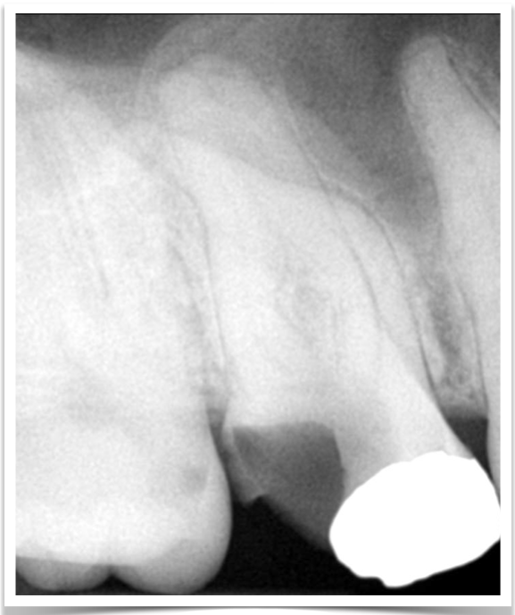
Clinical examination demonstrates a complicated fracture (fracture of enamel and dentin with pulpal exposure) and caries. The tooth responded normally to cold test and percussion. Probing and mobility were normal.
The radiological examination demonstrates the extent of the fracture. The mesial and distal roots were severely curved. The X-rays also show long roots.
Diagnosis
Normal pulp and normal periapical tissues.
TREATMENT
The treatment options offered to the patient were:
- Root Canal Treatment.
- Extraction
Treatment selection
The patient wanted to save his tooth and consented to do the root canal treatment.
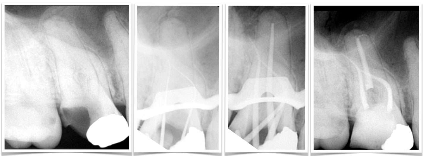
From left to right: 1- Pre-Operative X-ray. 2- MB & DB film check. 3- Cone Fit. 4- Final X-ray
Potential complications
The following potential complications were discussed with the patient:
- Perforation especially at the level of curvature
- File separation
- Ledge and blockage of the canals
Execution
- Working lengths were found with #6 hand files and confirmed with # 10 hand files
- A glide path was created using Scout Files
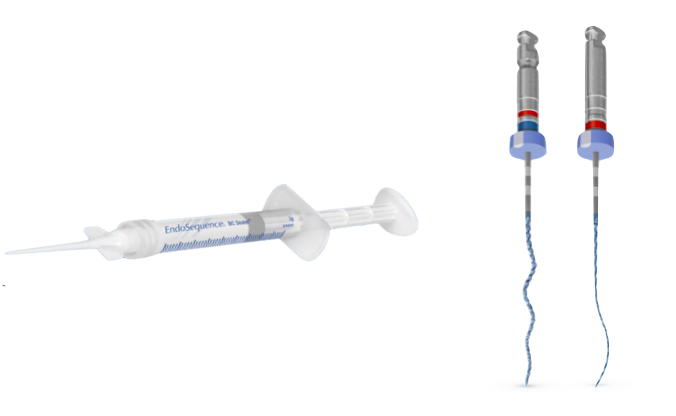
- Instrumentation with XP-3D Shaper and Finisher files
- Root canal filling with Bioceramic Sealer using cold hydraulic condensation
The 4 canals were 25mm in length.
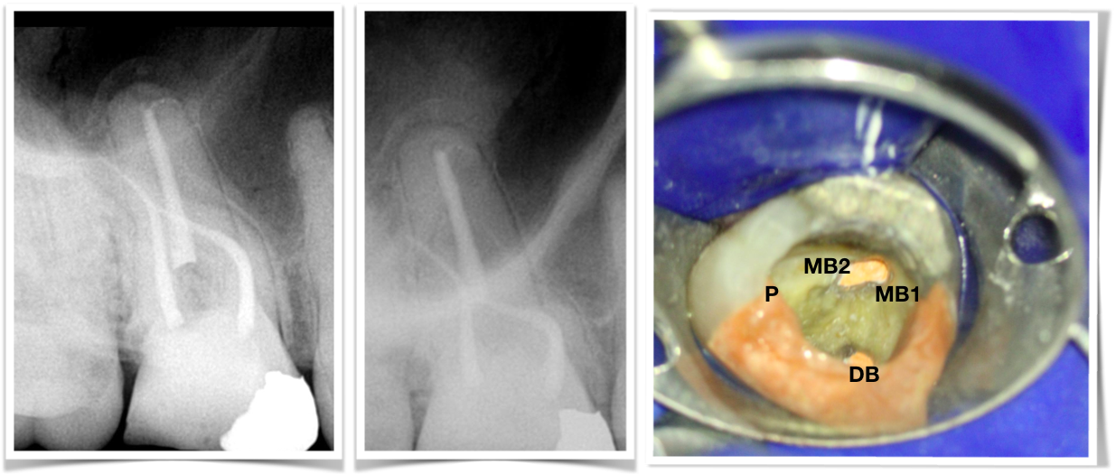
From left to right: 1- Final X-Ray. 2- Angulated view. 3- Clinical picture
More Information
How to manage curved root
- Ensure adequate access cavity
- Ensure proper glide path
- Gradual taper increase- Start with 2% then increase to 4%
- Gradual file size increase- Start with #6 hand files then increase to #8, #10 etc…
- Use ‘’ Scout Files ‘’ or ‘’ Path Files ‘’ for gradual increase in taper and apical diameter