A case study by Dr. Chafic Safi, Centre Endodontique St-Laurent
A 44-year-old male patient presented to our clinic with the following chief complaint:
“Doc, I need a root canal. My bottom tooth really hurts !!”
Analysis
Clinical exam shows a crown on tooth 47.
The tooth tested positive to percussion and negative to cold.
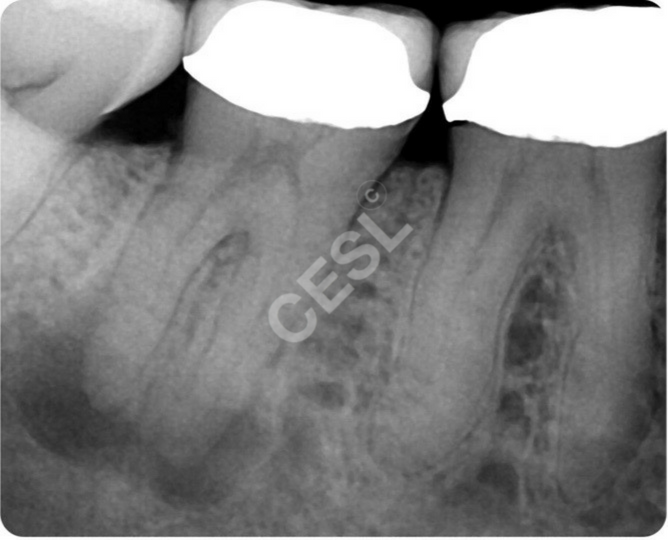
Radiological exam shows a tooth with 2 periapical radiolucencies and canal calcifications. The roots of the tooth were very close to each other and fused suggesting a particular configuration.
The other teeth in the area tested normal.

Diagnosis
The diagnosis was a Necrotic Pulp and Symptomatic Apical Periodontitis.
Treatment
The treatment options offered to the patient were:
- A root canal treatment
- An extraction
Treatment Selection
The patient opted for saving her tooth via root canal treatment.
Potential Complications
The following potential complications were discussed with the patient:
- Crown loosening
- Separation of an instrument
- Blockage/ledge
- Strip- perforation of the canals
The Treatment
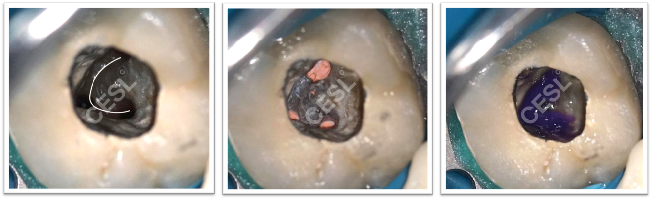
Retreatment was completed with cleaning of the canals in 3 dimensions using the XP-3D shaper and XP-3D Finisher files. Root canal obturation was achieved using Bioceramic Sealer and Bioceramic Gutta-Percha. The access cavity was sealed with a resin composite. The orifices were sealed with a purple flowable composite to ensure a flight-tight seal. The root canal system was in a shape of a ’’C’’. This configuration represents many challenges mainly the ability to clean the canals safely without weakening or perforating them. A maximum Apical Size of #30 is recommended in these cases.

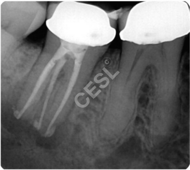
Left to right: 1- Biomechanical instrumentation completed. We can appreciate the ’’C’’ shaped system. 2- Root canal filling completed. 3- Orifice sealing with a flowable purple composite.