A case study by Dr. Chafic Safi, Centre Endodontique St-Laurent
A 75-year old male presented themselves to our office with a chief complaint:
“My dentist sent me for a root canal. I have calcified canals.”
Analysis
Clinical examination revealed the presence of an extensive bridge, with tooth #24 serving as an abutment. Tooth #24 responded negatively to cold and EPT and was tender to percussion.
Radiological examination revealed the presence of calcified canals and an apical radiolucency.
Diagnosis
The diagnosis was: Pulpal necrosis and symptomatic apical periodontitis..
Treatment
The treatment options offered to the patient were:
- Root canal treatment. Prognosis: Favorable.
- Extraction. Prognosis: Favorable.
Treatment Selection
The patient consented for root canal treatment.
The treatment
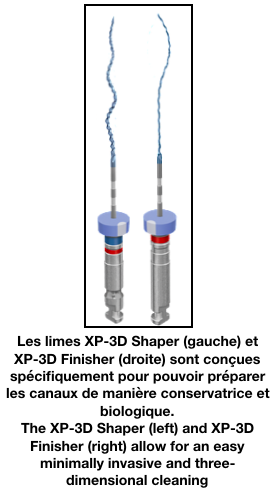
The procedure was completed in 2 visits with placement of Calcium Hydroxide as an intra-canal medicament. During the first visit, biomechanical instrumentation of the root canal system was completed using the XP-3D shaper and finisher files.
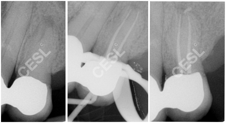
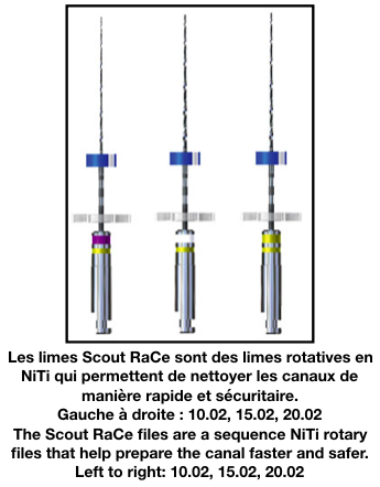
There were 2 canals in the tooth. Scout RaCe files were used prior to the XP-3D in order to ensure a proper glide path. Calcium Hydroxide was placed as an interim medicament and the tooth sealed with a temporary restoration.
Treatment was completed 10 days later. The canals were obturated using Bioceramic Sealer and Bioceramic Gutta Percha, offering superior biologic sealing and biocompatibility. The access cavity was sealed with resin composite.