A case study by Dr. Chafic Safi, Centre Endodontique St-Laurent
A 25 year-old male presented to our office with a chief complaint:
“I am not sleeping, I have a really bad toothache”
Analysis
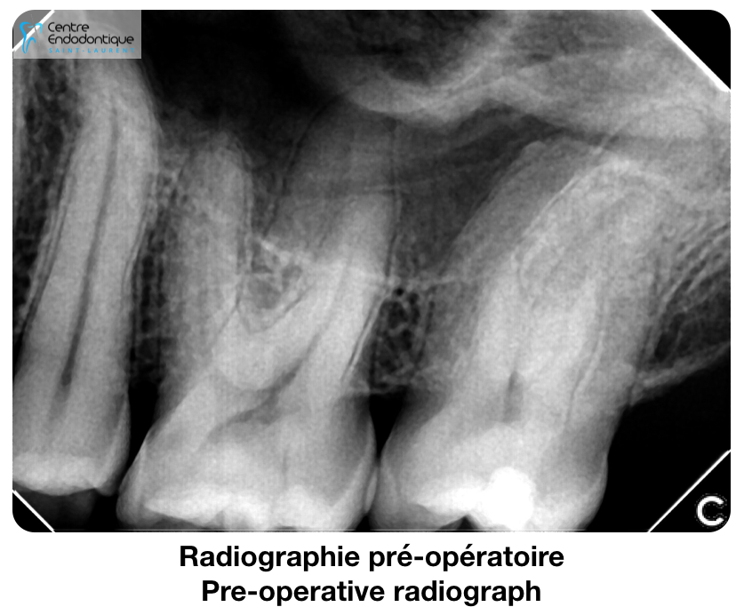
Clinical examination revealed the presence of a restoration on tooth #27. The tooth was painful to percussion. Cold and vitality tests were negative.
Radiological examination revealed the presence of a widened PDL around the palatal root of tooth #27.
Diagnosis
The diagnosis was: Pulpal necrosis and symptomatic apical periodontitis.
Treatment
The treatment options offered to the patient were:
- Root canal treatment. Prognosis: Favorable
- Extraction. Prognosis: Favorable
Treatment Selection
The patient consented to a root canal treatment.
The treatment
The procedure was completed in 2 visits with placement of Calcium Hydroxide as an intra-canal medicament.
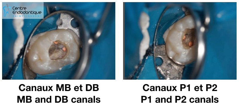
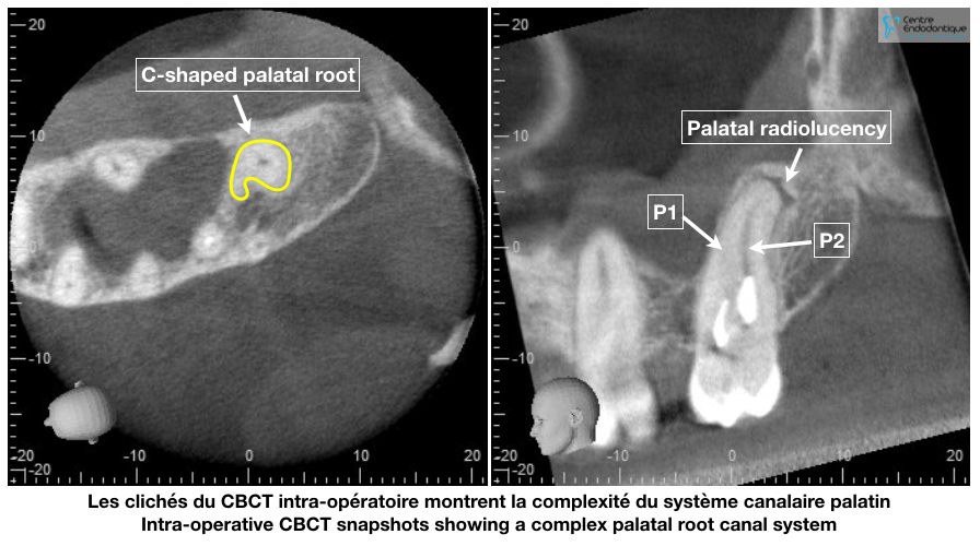
During the first visit, a special anatomy was discovered: the presence of 2 palatal canals in the tooth. This prompted us to take a 3D scan (CBCT), which luckily could be performed on site in our clinic.
CBCT snapshots revealed the presence of a C-shaped palatal system with 2 separate canals. Studies report the prevalence of such anatomy to be 0.4-1.3%.
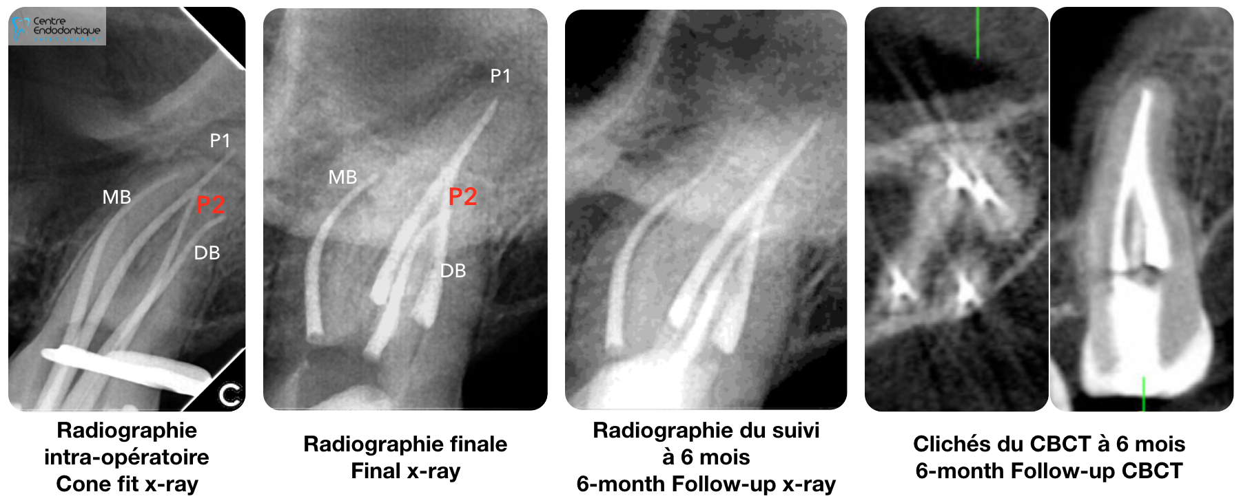
Treatment was completed 10 days later.
The final irrigation protocol for both visits consisted of the following: 6% NaOCl followed by 17% EDTA which was then followed by 2% Chlorhexidine.
All 4 canals, Palatal 1, Palatal 2, Disto-Buccal, and Mesio-Buccal were instrumented using the XP-3D Shaper file and obturated using cold hydraulic condensation with Bioceramic Sealer.
Follow-up
At 6-months follow-up, complete healing was observed on X-ray and on CBCT.