A case study by Dr. Chafic Safi, Centre Endodontique St-Laurent
A 55-year-old Caucasian female patient presented herself at our clinic with the following primary complaint:
“I can’t chew on my tooth.”
Analysis
History of chief complaint revealed that the patient started to develop symptoms upon chewing a couple of weeks ago. She consulted her dentist who referred her to our office.
Her chief complaint was reproduced upon percussion of her lower right first molar (#46).
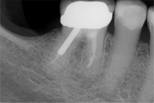
Clinical and conventional radiological exam further revealed that the tooth has a crown, a post, a root canal treatment, and a J-shaped periapical radiolucency around the mesial root. The periapical image also suggests a possible missed canal in the mesial root.
Probing and mobility were within normal limits.
Diagnosis
The diagnosis was a previously treated tooth and symptomatic apical periodontitis.
-
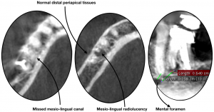
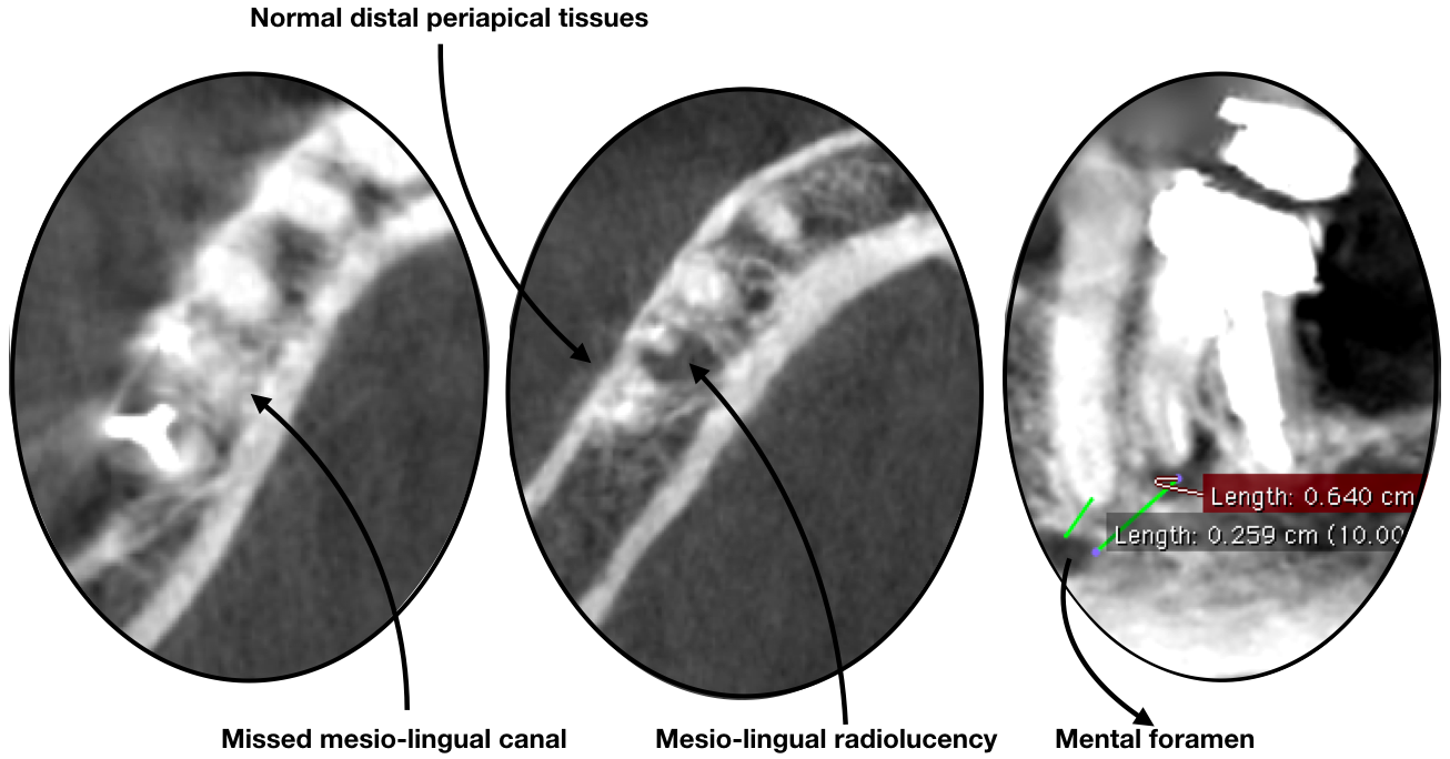
- Figure 1 (eng): L’imagerie en 3D du canal mésio-lingual manqué, tissus périapicaux normaux au distal, Foramen mentionnier / CBCT imaging of Missed mesio-lingual canal, normal distal periapical tissues, mesio-lingual radiolucency, and mental foramen
-

- Figure 1 (fr): L’imagerie en 3D du canal mésio-lingual manqué, tissus périapicaux normaux au distal, Foramen mentionnier / CBCT imaging of Missed mesio-lingual canal, normal distal periapical tissues, mesio-lingual radiolucency, and mental foramen

Treatment
The treatment options offered to the patient were:
- Endodontic microsurgery (commonly known as apicoectomy). Prognosis: Favorable
- Non-surgical root canal retreatment. Prognosis: Guarded
- Extraction. Prognosis: Favorable
Treatment Selection
The patient opted for an endodontic microsurgery.
Potential Complications
The following potential complications were discussed with the patient:
- Re-infection of the periapical tissues
- Possible damage to surrounding teeth
- Local paresthesia of the lower right lip and chin, which is why a CBCT was taken to assess proximity of the mental nerve
- Possibility of cracked tooth as suggested by J-shaped lesion
The Treatment
CBCT imaging confirmed the following:
- Absence of radiolucency on the distal root
- Missed mesio-lingual canal
- Safe distance form the mental foramen
Endodontic microsurgery was performed using magnification and illumination. 3 mm of the mesial root were resected using a Lindemann burr. After ultrasonic retro-preparation of both mesial canals, Bioceramic Root Repair Material was used to fill and seal the root canal space. No sign of cracked tooth or root fracture was observed.
Follow-up
A 6-month follow-up radiograph shows complete healing with reformation of normal periodontal space and lamina dura around the mesial root. The patient is asymptomatic, the crown is still in place and the tooth is saved.
No signs of paresthesia were ever felt.
More information
Advantages of Endodontic Microsurgery
- Safe and conservative approach
- High success rate
Additional Images
-

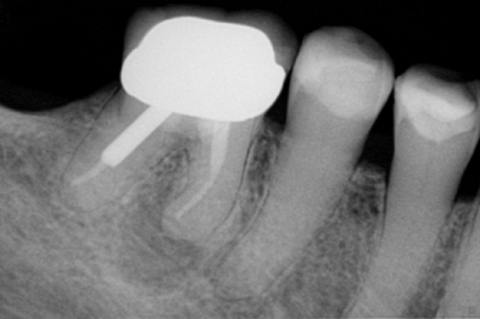
- Figure 2: Radiographie pré-opératoire / Pre-operative radiograph
-
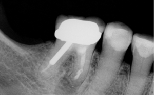
- Figure 3: Radiographie finale / Post-operative radiograph

-
- Figure 4: Radiographie du suivi au 6ème mois / 6-month follow-up radiograph
